Learn OCT Machine Basics: Facts, Details, and Practical Suggestions
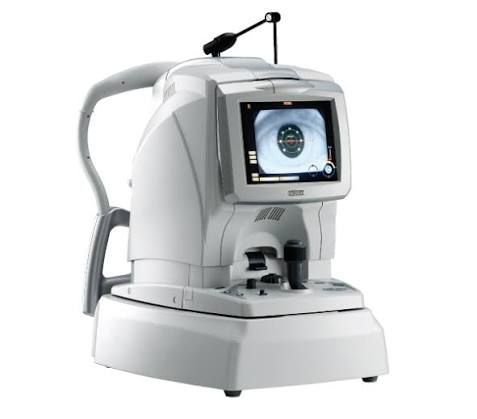
An OCT machine, or Optical Coherence Tomography system, is a medical imaging device that uses light to create detailed cross-sectional images of tissue. It is particularly well known in ophthalmology, where it can display different layers of the retina and help clinicians evaluate structural changes.
Unlike conventional photography, which generally produces a surface image, OCT imaging can provide depth-resolved information. This makes it useful for examining structures that cannot be evaluated adequately from a simple external photograph.
The technology is based on low-coherence interferometry. In simplified terms, the OCT system sends light into tissue and analyzes reflected light to estimate the location of different tissue structures. The system then converts this information into an image.
An OCT scan is generally non-invasive. In ophthalmic applications, the patient typically looks toward a fixation target while the equipment captures images. The procedure does not require the OCT system itself to physically contact the eye.
OCT is widely associated with retinal imaging, but different configurations can examine other anatomical structures. These include the anterior segment of the eye and, in specialized applications, cardiovascular structures.
How an OCT Machine Works
An OCT system combines optical components, scanning mechanisms, detectors, computing hardware, and image-processing software.
A simplified imaging process includes:
A light source generates suitable low-coherence light.
Optical components direct the light toward the tissue.
Reflected or backscattered light is collected.
Interference patterns are measured by the system.
Software processes the measurements into cross-sectional images.
The resulting images can be reviewed and compared over time.
Different OCT technologies use different acquisition and processing approaches. Time-domain OCT was an earlier approach, while spectral-domain OCT and swept-source OCT provide faster or deeper imaging capabilities in many modern systems.
The resulting images may be presented as two-dimensional cross-sections, three-dimensional volumes, thickness maps, or other quantitative displays.
Common Types of OCT Equipment
OCT equipment varies according to the anatomical area being examined and the intended clinical application.
| OCT type | General application |
|---|---|
| Retinal OCT | Imaging retinal layers and related structures |
| Spectral-domain OCT | High-resolution retinal and ophthalmic imaging |
| Swept-source OCT | Deeper or wider-range imaging in selected applications |
| Anterior segment OCT | Imaging structures at the front of the eye |
| OCT angiography | Imaging retinal and choroidal microvasculature |
| Intravascular OCT | Imaging structures inside blood vessels |
| Multimodal imaging systems | Combining OCT with other imaging techniques |
The exact capabilities of an OCT machine depend on its optical design, scanning range, detector technology, software, and intended use.
Why OCT Imaging Is Important
OCT imaging is important because it can provide detailed structural information without requiring tissue removal. In ophthalmology, this can help clinicians evaluate retinal and optic nerve structures.
The American Academy of Ophthalmology explains that OCT can show individual retinal layers and the optic nerve fiber layer. These measurements can assist in evaluating conditions such as glaucoma, age-related macular degeneration, and diabetic eye disease.
OCT does not by itself establish every diagnosis. Clinicians interpret OCT findings alongside symptoms, medical history, examination results, and other diagnostic information.
Common Uses of an OCT Scan
OCT scans are used in several areas of medical imaging.
Retinal assessment
Retinal OCT can show the thickness and structure of retinal layers. Changes in these structures can be relevant when evaluating or monitoring various retinal disorders.
Glaucoma assessment
OCT can measure structures such as the retinal nerve fiber layer and other regions associated with the optic nerve. These measurements can provide quantitative information that complements clinical examination.
Diabetic eye disease
OCT may help visualize retinal changes associated with diabetes. It can provide structural information that is useful for clinical assessment and monitoring.
Age-related macular degeneration
OCT is commonly used to examine the macula and monitor structural changes associated with retinal disease.
Anterior segment imaging
Specialized anterior segment OCT equipment can image structures at the front of the eye, including the cornea and other anatomical regions.
OCT angiography
OCT angiography, or OCT-A, uses motion contrast to visualize blood vessels in the retina and choroid. Unlike fluorescein angiography, it does not require an injected dye.
Factors That Influence OCT Image Quality
Good OCT imaging depends on more than the imaging device itself. Several factors can influence the resulting scan.
Important considerations include:
Patient fixation and movement.
Optical alignment.
Media clarity and ocular conditions.
Scan pattern and field of view.
Signal strength.
Segmentation accuracy.
Operator technique.
Software processing.
Appropriate interpretation.
Artifacts can appear in OCT images. For example, movement, blinking, poor fixation, shadows, or incorrect automated segmentation can affect the result.
For this reason, an OCT image should not be interpreted solely according to a single automated measurement. Reviewing the original scan and its quality is an important part of responsible clinical interpretation.
Recent Developments in OCT Technology
OCT technology continues to evolve through improvements in scanning speed, image resolution, software, multimodal imaging, and artificial intelligence.
OCT angiography expansion
OCT-A has become an increasingly important non-invasive imaging method for examining retinal and choroidal microvasculature. The American Academy of Ophthalmology published a beginner's guide to OCT-A on July 8, 2025, describing its use in depth-resolved vascular imaging.
New regulatory clearances in 2025–2026
Recent U.S. Food and Drug Administration records demonstrate continued development of OCT equipment. For example, the FDA recorded a substantially equivalent 510(k) decision for the SPECTRALIS HRA+OCT and related variants on May 7, 2026.
Another FDA record shows a March 13, 2026 decision involving the Pentacam Cornea OCT, an anterior-segment imaging device.
The FDA also recorded an April 24, 2026 decision involving an OCT imaging system used in cardiovascular imaging.
These regulatory records illustrate that OCT is not limited to one application area and that imaging technologies continue to develop across ophthalmic and cardiovascular fields.
Artificial intelligence and automated analysis
AI-based image analysis is another important research and technology direction. Algorithms can assist with image classification, segmentation, measurements, and detection of patterns.
However, automated analysis should be evaluated carefully because performance can vary according to image quality, patient population, disease characteristics, and the dataset used to develop the algorithm.
OCT Machine Regulations and Policies in India
OCT equipment used for medical purposes falls within India's medical-device regulatory framework. The Central Drugs Standard Control Organization, or CDSCO, maintains the regulatory framework and publishes information concerning medical devices and their classification.
India's Medical Devices Rules, 2017 provide the regulatory structure for medical devices, including requirements relating to classification, manufacturing, importing, licensing, and quality-related processes. CDSCO continues to publish amendments, guidance documents, and classification information.
A CDSCO classification document identifies an optical coherence tomography imaging system as a Class C medical device in the referenced classification list. The description covers diagnostic imaging of tissue characteristics using near-infrared light.
For specialized cardiovascular OCT, a CDSCO risk-classification document published in November 2025 lists a coronary optical coherence tomography system catheter as a Class D device.
Regulatory classification can depend on the intended use and device configuration. Manufacturers, importers, healthcare institutions, and other regulated entities should therefore consult current CDSCO notifications and applicable requirements rather than relying on a general classification summary.
Useful OCT Tools and Educational Resources
Understanding OCT is easier when learners use reliable medical and regulatory resources.
American Academy of Ophthalmology: Provides educational material about OCT, OCT-A, retinal layers, and clinical interpretation.
CDSCO: Provides Indian medical-device regulations, classification documents, guidance, and regulatory updates.
U.S. FDA: Its medical-device database provides regulatory records for cleared OCT devices in the United States.
OCT image atlases: Academic ophthalmology resources can help learners compare normal and abnormal retinal structures.
Clinical imaging software: OCT systems commonly include software for viewing scans, measuring retinal thickness, creating maps, and comparing examinations.
Image-quality checklists: Structured checklists can help users review fixation, signal quality, segmentation, artifacts, and scan coverage.
For educational purposes, learners should distinguish between an OCT image used for teaching and a clinical diagnostic interpretation performed by a qualified healthcare professional.
Practical Suggestions for Understanding an OCT Scan
When reviewing an OCT image for educational purposes, it can be useful to follow a consistent sequence:
Check whether the scan has adequate image quality.
Identify the anatomical region being displayed.
Become familiar with normal retinal or tissue layers.
Look for obvious changes in structure or thickness.
Review automated segmentation lines for accuracy.
Compare measurements with appropriate reference information.
Consider whether artifacts could affect the appearance.
Avoid drawing a medical conclusion from a single image without clinical context.
The American Academy of Ophthalmology's 2026 OCT educational material emphasizes first becoming familiar with normal retinal anatomy before interpreting abnormal patterns.
Frequently Asked Questions
What is an OCT machine?
An OCT machine is an optical imaging device that uses light and interferometric measurement techniques to create cross-sectional images of tissue. It is widely used for retinal and ophthalmic imaging.
Is an OCT scan invasive?
Ophthalmic OCT is generally a non-invasive imaging examination. It uses light to obtain images and normally does not require contact with the eye or an injected contrast agent.
What can an OCT scan show?
Depending on the system and application, an OCT scan can show tissue layers, thickness measurements, structural changes, and other anatomical features. Retinal OCT commonly displays detailed retinal layers.
What is the difference between OCT and OCT angiography?
Standard OCT primarily provides structural information. OCT angiography uses motion-based optical signals to visualize blood flow patterns in retinal and choroidal vessels without requiring dye injection.
Can an OCT machine diagnose an eye condition by itself?
No. OCT provides imaging information, but diagnosis generally requires interpretation by a qualified healthcare professional together with examination findings, medical history, symptoms, and other relevant tests.
Conclusion
OCT machines have become important tools in modern optical imaging because they can provide detailed cross-sectional information about tissue without requiring invasive tissue sampling. In ophthalmology, OCT imaging is particularly useful for examining retinal layers, the optic nerve, and other eye structures.
Understanding the basics of Optical coherence tomography, OCT equipment, OCT scans, and OCT angiography helps students and general readers understand how modern medical imaging works. At the same time, responsible interpretation requires attention to image quality, artifacts, clinical context, and the limitations of automated analysis.
Recent developments in OCT include expanding OCT angiography applications, improvements in imaging systems, multimodal technologies, and growing use of computational analysis. As these technologies continue to develop, appropriate regulation, clinical validation, data quality, and professional interpretation remain important parts of safe and effective medical imaging.

Related Post...

Explore Secure Checkout: A Complete Guide to Payment Security and Protection
Updated: November 06, 2026
Read More
Learn the Basics of Choosing a Degree: Facts, Details, and Practical Suggestions
Updated: November 06, 2026
Read More
Learn Multi-Layer Pantry Shelf Organizer: Essential Basic, Fact, and Useful Knowledge
Updated: November 04, 2026
Read More